RADIATION THERAPY FACILITIES
Facilities in Radiation Therapy
The Radiation Oncology Department at RGCIRC has facilities for both
- External Beam Radiotherapy (EBRT) and
- Brachytherapy
External Beam Radiation Therapy
The external beam radiation therapy is the form of radiation treatment, delivering several beams of high-energy photons, electrons or protons through the skin to the cancer site and the immediate surrounding area in order to destroy the cancer cells. Typically the patient lies on a couch and the Gantry or Head of the machine rotates around, delivering radiation at a particular part of the body.
While megavoltage photons or protons are used to treat more deep-seated cancers (e.g. bladder, bowl, prostate, lung or brain), Electron beams are used for treating skin cancer and cancers seated in superficial tissues.
Steps in Radiation Treatment
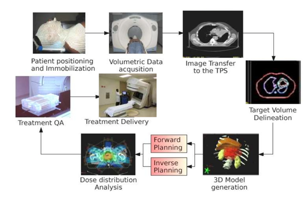
Simulator
Simulation is the most important preparation step before the actual radiation treatment.
Siemens Somatom Sensation-Open CT- Simulator —The simulation room is equipped with this dedicated large-bore CT scanner called as Simulator.
During Simulation,–
- The treatment setup is replicated by positioning the patient on the flat couch immobilized by specially designed devices.
- The patient is then aligned with the help of reference low-energy lasers in the room and marked on the skin with tattoos.
- Finally, a CT scan is performed to acquire the anatomy of the reference treatment site.
- This CT scan would be used to identify the tumour area and also the surrounding normal critical organs to create a treatment plan that will guide the treatment machine to target the tumour accurately and spare the normal organs.
- The simulated setup will be exactly reproduced before each treatment by matching the reference lasers in the treatment room to the tattoos and comparing the 2D/3D on-board images with the simulation CT scan. The patient may need to spend from 15 minutes to 45 minutes to complete the entire process of simulation.
The department also has a 2D Simulator — Varian – Acuity Simulator, which is a specialized X-Ray fluoroscopy unit, having the same parameters as those of the therapy unit without its treatment capabilities.
A change of planning during the course of the treatment may often be need after a few days of radiation for selected cases using this equipment .
Linear Accelerator, or Linac
This is a machine that is commonly used to deliver external beam radiation treatments ( EBRT) to cancer patients. A linear accelerator is programmed to deliver high-energy X-rays that conform to the specific size, shape and location of a tumor. In this way, the LINAC can target and destroy cancerous cells in a precise area of a patient’s body with minimal exposure to the surrounding healthy tissue.
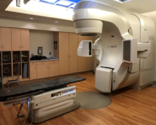
The Radiation Oncology Department is equipped with 4 LINACs including , Accuray- Tomotherapy Radixact X9 with 6FFF photon beam with MV Imaging, capable of advanced state of arts IMRT, IGRT, Volumetric therapy and TBI techniques, Varian – Clinac-iX Rapid Arc, Varian – True Beam Stx, Elekta -Synergy with facilities of Photon Beam, Electron Beam, providing
- 3D-Conformal Radiotherapy
- Intensity Modulated Radiation Therapy – IMRT
- Image Guided Radiotherapy (IGRT)
- Volumetric Modulated Arc Therapy (VMAT)
- Stereotactic Radiotherapy (SBRT)
- Tomotherapy
- Brachytherapy


