DIAGNOSTIC AND THERANOSTIC MARKERS


Overview
The department of surgical pathology includes the sections of Histopathology, Cytopathology and Immunohistochemistry. The department’s strength is its highly experienced team of Oncopathologists. The quality of reporting is further enhanced by the fact the cases are opined by two oncopathologists, one of them being site-specific specialist. The site specific practice has helped department to deliver accurate and clinically useful reports to our clients. The faculty is an active member in all multispecialty clinics, contributing immensely in the diagnosis, predictive and prognostic outcomes, hence providing comprehensive patient care. In addition the entire department is involved in research work.
The Immunohistochemistry section, the backbone of our department, is one of the leaders in Northern India with more than 140 theranostic and diagnostic markers. The department is early adopter of new clinical relevant test like ALK-1 & PDL-1 immunoassays. At present our biomarker testing complements molecular testing to include MMR protein, P16, ATRX, IDH-1. All validated markers are used for routine reporting on fully automated immunostainers.
The technical staff has been well trained for tissue conservation practices, delivering good quality sections, complimenting H & E with special stains. The rapid intra-operative diagnosis and margin assessment is provided in-house and to neighboring hospitals, by frozen section.
The gross photographs are captured by Macro path gross imaging system and stored in hospital PACS for future reference and archiving.
Men being most important, machines also are needed to facilitate the optimization of efforts and outcomes. In this regard the laboratory has the best and the latest equipments with backups to ensure fastest TAT at all times. Vantage workflow system ensures minimum transcription errors; mix up of blocks and follow-up ordering in smoothened & glitch free manner.
The cytopathology section receives body fluids, FNAC material, and exfoliative cytology, including gynecological smears. The department uses liquid based cytology and cell block preparation for optimizing the diagnostic yield and aid in ancillary techniques. The rapid onsite screening has been practiced for all FNA procedures performed by pathologist and is also provided during guided procedures.
To keep the analytical process of the department quality at par with recommended international benchmarks, Lab regularly participates in proficiency testing (NordiQC, UKNEQAS etc.)
The department has an established DNB training program, fellowship in oncopathology & DMLT training program with an extensive and elaborate teaching curriculum with participation of the entire department.
Immunohistochemistry
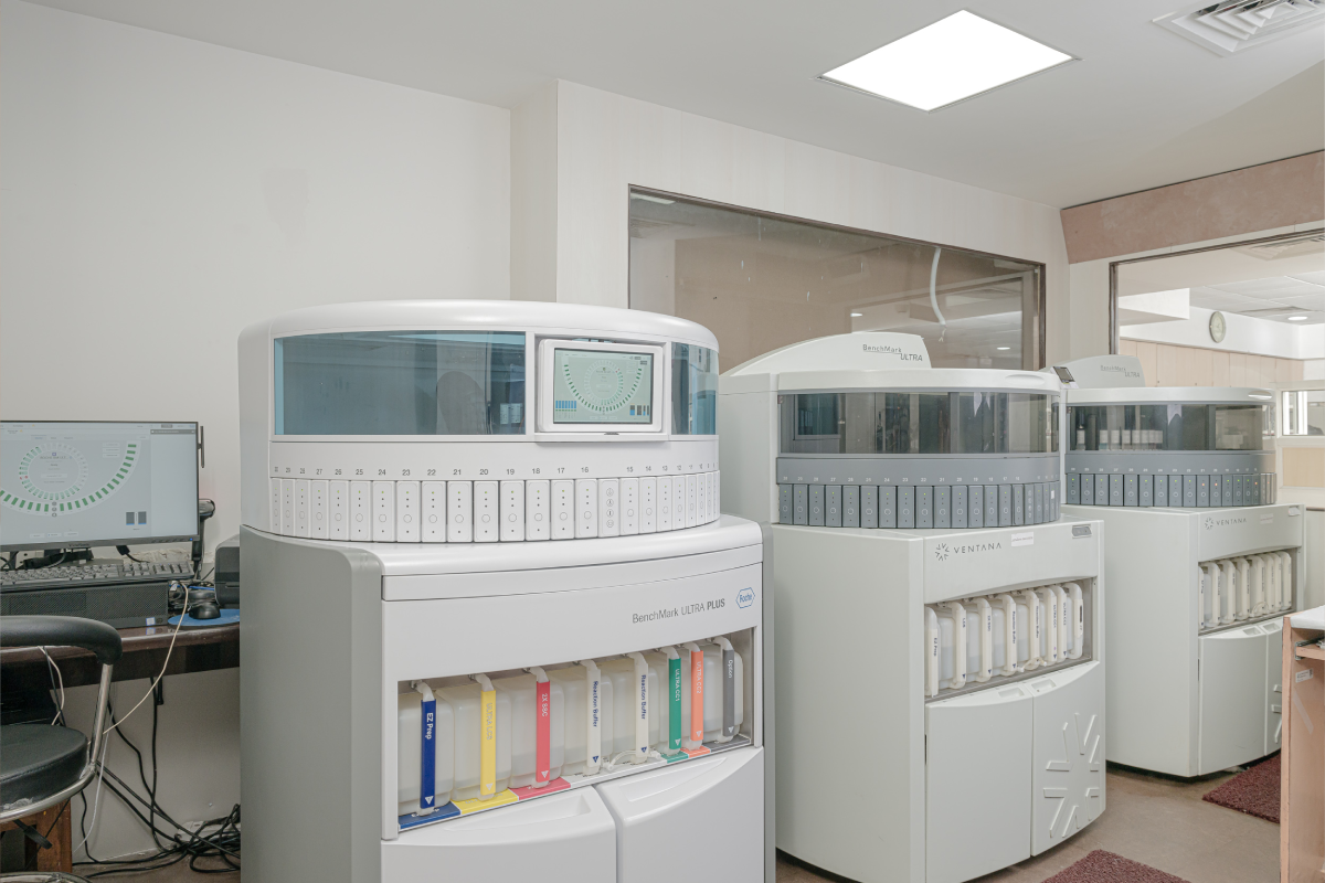
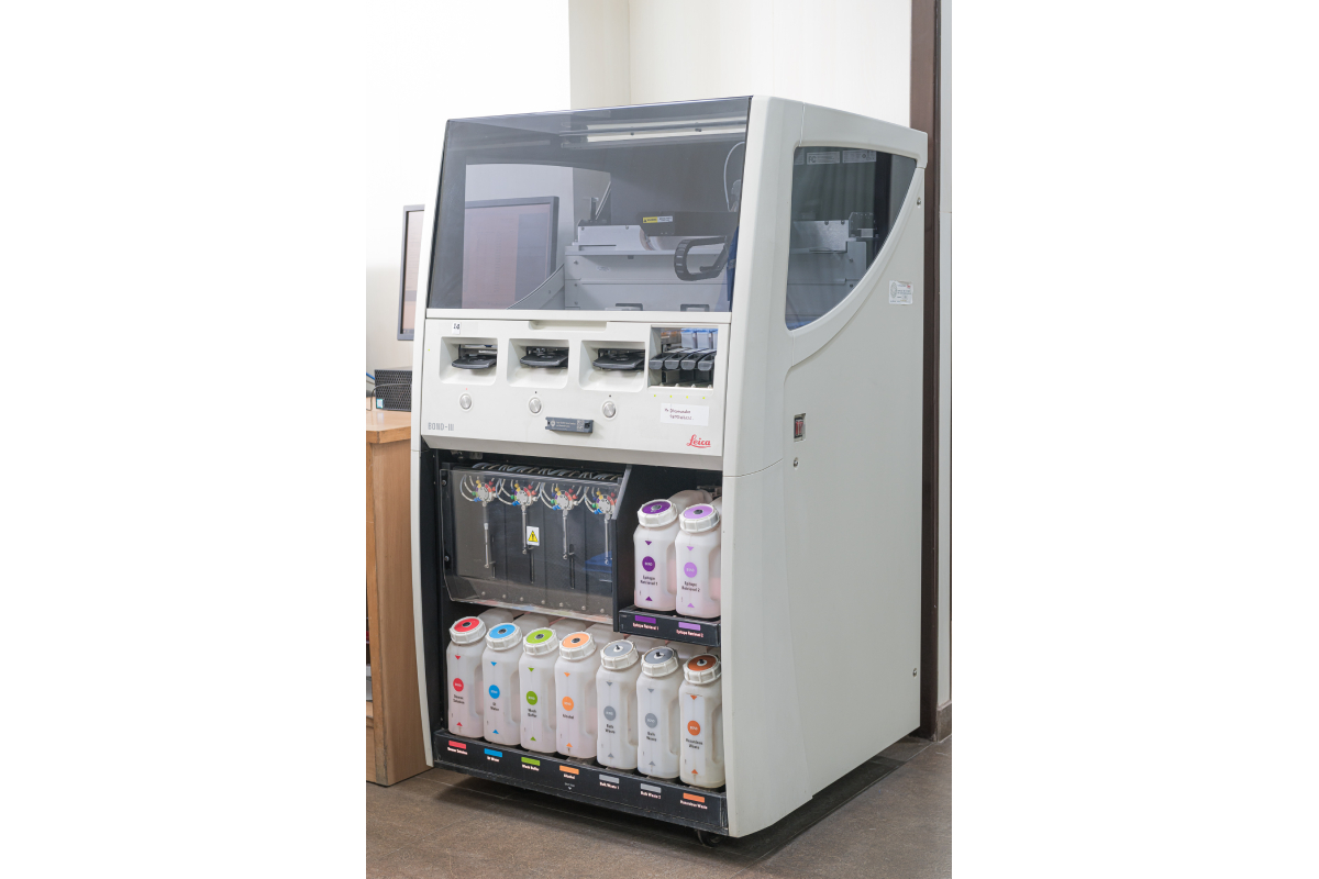
Immunohistochemistry (IHC) has evolved and expanded its use in pathology from diagnosis and classification, to predictive and prognostic utility. Our laboratory provides a wide range of these markers.We have two state of the art latest fully-automated IHC platforms,VentanaBenchMarkXT, VentanaBenchMark Ultra (https://diagnostics.roche.com/global/en/products/instruments/benchmark-ultra.html) and Leica Bond III systems.
- Bone and Soft tissue: H3F3A (Histone 3.3) G34W is used as a surrogate marker for H3.3 mutations, which helps in confirming primary benign and malignant giant cell tumor (GCT) of bone, and distinguish it from other mimics like giant cell osteosarcoma, with major therapeutic implications. HHV-8(Human herpesvirus-8), is available for diagnosing Kaposi sarcoma.H3K27me3is available for diagnosingmalignant peripheral nerve sheath tumors (high grade and radiation induced MPNST), which suggests loss of PRC2 pathway as the underlying molecular mechanism. Specific markersCDK4 & MDM-2 are available for diagnosing parosteal osteosarcoma (OS), intramedullary low grade OS,and liposarcoma (well differentiated & de-differentiated liposarcomas).NKX2.2 is a sensitive and specific marker for diagnosing Ewings’ sarcoma, and forms a target of EWS-FLI-1 fusion protein.INI-1/ SMARCB1is done for diagnosing the aggressiveextrarenalrhabdoid tumors.
- Breast: Specific markersGATA-3 and Mammaglobin help in confirmation of breast origin, especially in metastatic sites. Androgen Receptor (AR)immunoexpression is also being done triple negative breast cancer (TNBC).
- Central nervous System (CNS)/Brain: Biomarkers in CNS tumors assist the diagnosis, prognosis, and predict therapeutic response. IHC performed at our laboratory include ATRX, IDH1 (R132H), TP53 and BRAF V600E, which assist in classifying glial tumors.Medulloblastomas are classified into appropriate genetic groups by beta-catenin, YAP1, and GAB1, and p53.Ependymoma specific markers include L1CAM and YAP1. H3K27M, used to classify high grade midline gliomas, is also available in our armamentarium. INI1/SMARCB1is used to identify atypical teratoid/rhabdoid tumors (AT/RT), which have a significantly different treatment regime from other CNS embryonal malignancies.
- Female genital tract: A panel of markers is required for diagnosis and characterization of cervix, endometrial and ovarian carcinomas (at the primary and metastatic site) including PAX-8, ER, p53, p16, WT-1 and Napsin-A. L1CAM and HER2 are the upcoming markers used for prognostication of endometrial carcinomas. IHC for mismatch repair (MMR) proteins (discussed later) are performed in endometrioid adenocarcinomas to identify cases with Lynch Syndrome. p16 is routinely performed in cervical carcinomas as a surrogate marker for HPV (human papilloma virus) association.
- Gastrointestinal tract: Periampullary carcinomas: Panel of markers including for CK20, CDX2, MUC1 and MUC2 is used to subclassifyperiampullary carcinomas into pancreatobiliary and intestinal type adenocarcinomas. SMAD-4 (DPC4) gene is done in pancreatic carcinomas.
- Head and Neck Squamous cell carcinomas: P16is used as a surrogate marker for HPV associated oropharyngeal carcinomas.NUT is available for aggressive NUT midline carcinomas
- Lymphomas: Apart from the routinemarkers for diagnosing lymphoproliferative disorders and lymphomas, we have updated our armamentarium with new markers with diagnostic and prognostic utility- LMO2, MNDA, TBET, GATA3, c-myc, EBER-ISH (in-situ-hybridization), and HHV-8(for castleman disease).
- Neuroendcrine Tumors: INSM-1 (Insulinoma associated protein-1) is a new marker for neuroendocrine differentiation (along with synaptophysin, chromogranin and CD56). INSM-1 is a reliable, sensitive and highly specific marker for neuroendocrine differentiation in tumors of various organs.
- Prostate: NKX3.1 is an androgen regulated prostatic tumor suppressor gene, and staining for protein is performed for diagnosing prostatic origin, especially at the metastatic sites. It is a highly sensitive and specific marker.
- Renal Tumors: A panel of markers isperformed for confirmation and subtyping of renal cell carcinomas (RCC) using CA-IX, CD10, CK7, and AMACR.SDH-B is performed to identify cases SDH-B deficient RCC. 2-SC (2-succinocysteine) and FHis available to identify Fumaratehydratase (FH) deficient RCC which are aggressive hereditary tumors; and TFE-3 and TFE-Bfor Mit Family of RCC.
- Thoracic tumors: BRG-1 (SMARCA4)is used as a prognostic marker in lung malignancies.
Theranostic and Prognostic Biomarkers
Discipline of immunohistochemistry for the surgical pathologist has been evolving rapidly, and it also has theranostic and genomic applications. IHC has been adapted to the identification and demonstration of both prognostic and predictive markers.
The term “theranostics” is used to describe the proposed process of diagnostic therapy for individual patients and to tailor a treatment for them based on a test result.
Hormonal Receptors [Estrogen Receptor (ER),Progesterone Receptor (PR)]
Breast Carcinomas– Hormone receptors {Estrogen receptor (ER) and Progesterone receptor (PR)} and HER2 testing is recommended to be done on all primary invasive breast carcinomas and on recurrent or metastatic tumors, and reported as per guidelines published by the American Society of Clinical Oncology (ASCO) and the College of American Pathologists (CAP).
Hormone receptor (HR) expression in invasive breast carcinoma has both prognostic and predictive significance; the use of endocrine therapy in HR-positive breast carcinomas has been shown to reduce the rates of recurrence and mortality.
The recommended pre-analytic and analytic variables required for tests, like cold ischemia time, are strictly maintained.
Endometrial Carcinomas – Hormone receptor (ER and PR) expression is also assessed on primary invasive endometrial endometrioid adenocarcinomas to predict response to endocrine therapy.
Human epidermal growth factor receptor 2 (HER2) Receptor testing
Breast Carcinoma- HER2 status is primarily evaluated to determine patient eligibility for anti-HER2 therapy in breast carcinomas. Reflex FISH (fluorescence in situ hybridization) test is performed in equivocal cases to confirm the amplification status.
Female genital tract – HER2 Testing is done on high grade endometrioid and serous endometrial carcinomas as a prognostic marker.
Gastric carcinoma – Assessment for tumor HER2 overexpression using IHC is done to determine patient eligibility for anti-HER2 therapy.
Salivary gland tumors- HER2 expression is done using IHC as a diagnostic marker for the aggressive salivary duct carcinomas.
BRAF V600E
BRAF V600E is done as a surrogate marker for BRAF gene mutations (with predominant mutation seen at V600E). It has a role in thyroid papillary carcinoma, colon adenocarcinoma, and melanoma, with therapeutic implications.
Human papillomavirus (HPV)
P16 immunohistochemistry is used as a surrogate marker of active HPV in head and neck oropharyngeal squamous cell carcinomas (OPSCC), and cervical carcinomas.
Epstein-Barr virus (EBV) detection using in situ hybridization (ISH) for EBV-encoded small RNAs (EBER) in nasopharyngeal carcinomas, lymphoepithelial carcinomas, and also for several types of hematopoietic malignancies.
NUT (nuclear protein in testis) : NUT is used as a diagnostic marker for NUT midline carcinoma which is a rare, aggressive, squamous cell carcinoma variant uniquely defined by NUT gene translocations.
Succinate Dehydrogenase (SDH)
SDH-B is performed to identify cases with germline/sporadic mutation in the enzyme succinate dehydrogenase, seen in gastrointestinal tumors (GISTs), extra adrenal paragangliomas, pheochromocytomas and some renal tumors (SDH-B deficient RCC).
PD-L1 (programmed death ligand-1)
RGCIRC was the first centre in North India to introduce PD-L1 immunoexpression testing by IHC as a predictive marker of immunotherapy in various solid organ tumors. PD-L1 scoring is done by Roches’s Ventana SP263 clone, on Ventana Benchmark XT autostainer. The SP263 assay is CE (European conformity) labeled to inform treatment decisions in lung cancer patients being considered for Keytruda (Pembrolizumab) immunotherapy as a first line of treatment.
PDL-1 scoring for by monoclonal antibody SP142 is done on benchmark ultra Ventana autostainer, which is FDA approved assay for metastatic triple negative breast cancer (TNBC), Non-small cell lung carcinoma (NSCLC) and urothelial carcinoma for patient selection, to be treated with Tecentriq (atezolizumab).
Mismatch Repair (MMR) Immunohistochemistry Testing
IHC testing for DNA MMR protein expression (MLH1, MSH2, MSH6, and PMS2 expression) is performed to detect germline mutations in these MMR genes, and identify Lynch syndrome (hereditary nonpolyposis colorectal cancer syndrome [HNPCC]) which is associated with risk of cancers of colorectal, endometrial, gastric, upper urinary tract origin. HNPCC is an autosomal dominant inherited cancer syndrome with mutation in DNA mismatch repair genes. Loss of one or more proteins by IHC is suggestive of defective DNA mismatch repair within the tumor.