MOUTH & ORAL CANCER
Signs and Symptoms of Mouth & Oral Cancer
Globally, oral cancer is the sixth most common type of cancer and India contributes to almost one-third of total oral cancer cases. In India, around 80,000 new cases are reported annually.
Oral cavity includes the lips, the front two-thirds of the tongue, the gingiva (gums), the buccal mucosa (lining inside the cheeks and lips), the floor of the mouth, the hard palate (bony top of the mouth), and the small area behind the wisdom teeth. (Retromolar trigone)
Most common symptoms:
- Non-Healing Ulcer
- Bleeding
- Lump or swelling in the jaw, face, or mouth
- Loosening of teeth
- Pain, numbness, or weakness on the face
- Difficulty in opening the jaws and chewing
- Difficulty in swallowing
- Change in speech
- Pain in the ear or hearing loss
- Difficulty in breathing
- White or red patches in the mouth
- Loss of weight
- Swelling in the neck
Types of Mouth & Oral Cancer
- Lip Cancer
- Cheek/Buccal mucosa cancer
- Lower alveolus/Upper alveolus cancer
- RMT (Retromolar trigone) Cancer
- Floor of mouth
- Tongue
- Hard Palate
Majority of oral cancers are squamous cell carcinomas.
Screening and Diagnosis of Mouth & Oral Cancer
Always look at and in your mouth while brushing your teeth.Report any white or red patch or non healing ulcer in your mouth. Consult a dental surgeon annually as part of health check up.
Diagnosis of Oral Cancer
To find the cause of symptoms, a surgical oncologist evaluates a person’s medical history, performs a physical examination, and orders diagnostic tests. The examination and tests conducted may vary depending on the symptoms. Examination of a sample of tissue under the microscope is always necessary to confirm a diagnosis of cancer.
We insist on clinical examination and believe that a “Hand Scan is better than a CAT Scan”
Physical examination may include visual inspection of the oral and nasal cavities, neck, throat, and tongue using a small mirror and/or lights. The surgical oncologist may also feel for lumps in the neck and palpate lips, gums,tongue and cheeks.
Myth: Biopsy or FNAC leads to the spreading of cancer.
A biopsy means removal of a small piece of tissue. A pathologist studies the tissue under a microscope to make a diagnosis. A biopsy is the only sure way to tell whether a person has cancer.
- Endoscopy of relevant area wherever indicated.
- Laboratory tests examine samples of blood, urine, or cells from the nodes.
- X-rays create images of areas inside the head and neck on film.
- CT scan is a series of detailed pictures of areas inside the head and neck.
- CT scan is a series of detailed pictures of areas inside the head and neck.
- Magnetic resonance imaging (or MRI).
- PET scan in selected cases.
If the diagnosis of cancer is confirmed, we want to know the stage (or extent) of the disease. Staging is a careful attempt to find out whether cancer has spread and if so, to which parts of the body. Knowing the stage of the disease helps the surgical oncologist plan treatment.

Tumor Board/ Multispecialty Clinic Evaluation
Each and every Head & Neck cancer patient is evaluated by a special team of Surgical Oncologists (Head & Neck unit), Medical Oncologists, Radiation Oncologists, Onco-pathologists, and Imaging Specialists. Depending on the age, general condition,profession of patient, type of pathology, and stage of the disease, a custom-made treatment plan is charted out for each and every patient as per International Treatment Guidelines. (NCCN – National Comprehensive Cancer Network).
Treatment of Mouth & Oral Cancer
What do We Offer at RGCIRC?
- Composite Resection/ Commando operation for oral cancers
- Maxillectomy
- Neck dissections
- Skin lesions of face.
- Plastic Surgery: Reconstruction with Free flaps (Anterolateral Thigh flap, Medial Sural Artery Perforator flap, Radial Artery Forearm flap, Free Fibula flap), various local and regional flaps for reconstruction of face ,neck and oral defects.
Frozen Section
It is a modern technique to ensure the complete removal of cancer during surgery. The main cancer specimen is immediately sent to the laboratory after surgery for assessment by the Frozen section technique. The pathologist quickly freezes the sample from the margins of the specimen in a cryostat and takes a thin slice of the sample from various areas. They freeze the tissue at -15 to -20 degrees centigrade. They then place this under a microscope, analyze the sample, and report within 30 minutes.
Advanced Cancer of cheek excised and reconstruction done with free flap

Cancer of hard palate operated and rehabilitated with obturator


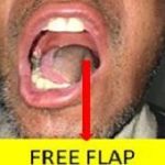
Cancer of tongue operated and reconstructed using free flap

Cancer of lower lip operated and reconstructed using free flap

Cancer of right lower jaw operated and reconstructed using free flap

Causes and Risk Factors of Mouth Oral Cancer

Tobacco-Smoking, Chewing

Gutakha Chewing

Alcohol

Sharp Teeth and Poor Orodental Hygiene

HPV Infections

Syphilis
DO’s after Surgery
- Speak less to ensure proper healing of stitches in the mouth and tongue.
- Liquid diet with adequate salt has to be given at regular intervals of 2 hours.
- Feeding through tube should be given with patient in sitting or 300 head-up position. Each feed of around 300 ml should be given slowly over a period of 20 minutes.
- All medicines as prescribed in the discharge note along with medicines for chronic diseases like diabetes, hypertension, thyroid-related, and asthma should be continued as earlier.
- Oral hygiene with Betadine mouth wash in 1:1 ratio with water to be done alternately with Hydroxyl mouth wash in 1:1 ratio with water every 2 hourly.
- In the case of free flap reconstruction patient should sleep in a straight position and follow instructions of plastic surgeon.
- Walking and moving around is a must.
- If the tracheostomy tube is there you may have to clean the inner cannula frequently preferably every 2-3 hourly.
- Keep the bib wet around the tracheostomy tube.
- Shoulder exercises as and when advised.
- Minimal blood-stained discharge from tracheostomy tube secretions is considered normal, don’t panic.
- Care of the drain and output charting as advised..
- Care of the wound as advised.
DON'Ts after Surgery
- Avoid sudden exertion to the head and neck area.
- Avoid room heaters if you have a tracheostomy tube.
- Avoid addictions.
- Avoid brushing your teeth.Follow instructions of your surgeon regarding diet,mouth care.
When to Contact the Treating Team?
- As per appointment.
- If the swelling around the neck wound or facial wound increases suddenly or within few hours.
- Bleeding from neck wound or oral cavity. Minimal blood-stained discharge from neck wound or during mouth wash needs attention.
- If there’s difficulty in breathing or swallowing, fever.
